Surgeons

YOUR IONM PARTNER

SURGEONS

Results from research published in peer-reviewed journals indicates that our monitoring services will significantly reduce the costs associated with surgically induced neurological deficits. By improving surgical outcomes and reducing short and long term costs, we can help you provide your patients with the most effective services available.
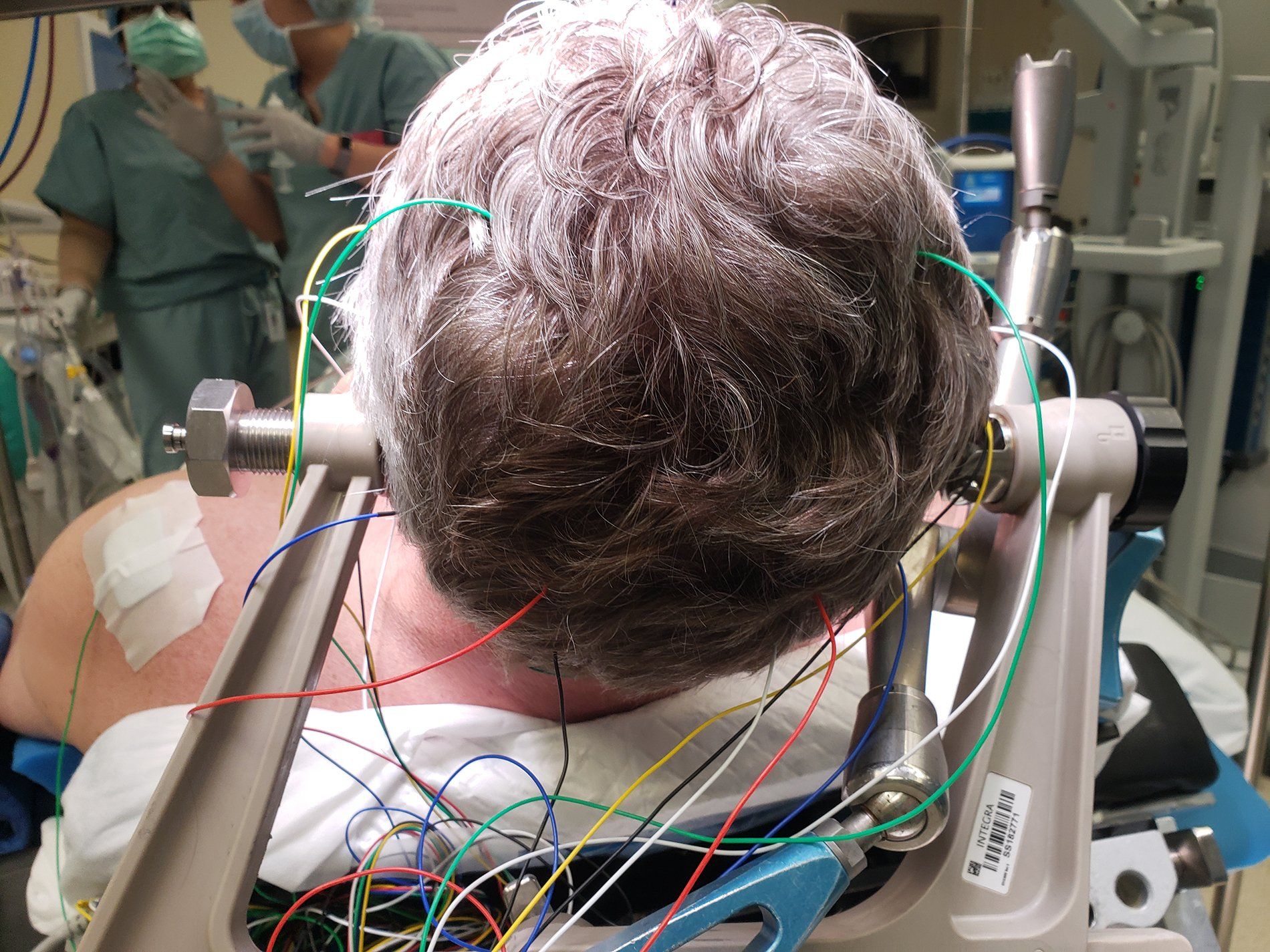
At The Phoenix, clinical excellence is one of our top priorities. All of our surgical neurophysiologists/technologists are board-certified or preparing for The Certificate in Neurophysiologic Intraoperative Monitoring (CNIM®), the industry-standard in intraoperative monitoring services. Using sophisticated and secure telemedicine services, all your cases will be monitored by our onsite technician and remotely by one of The Phoenix’s supervising neurologists.
We believe that trust and reliability are essential for a surgical team to operate efficiently, which is why we employ local technicians who are available on a 24-hour basis. The relationships that form out of this consistency lead to improved communication and patient safety.

Our privately held ownership allows us to cut through the corporate red tape and offer unparalleled customer service and clinical excellence from our local, skilled technicians.

PROCEDURE TYPES WE MONITOR

Below you will find the surgery types that we can monitor intraoperatively.

Orthopedic Surgeries

  • Spinal Fusion
  • Scoliosis Correction
  • Corpectomy
  • Discectomy
  • Laminectomy
  • Spinal Osteotomy
  • Acetabular Fractures and Revision 
050 - Human Brain Created with Sketch.

Neurological Surgeries

  • Cerebral Aneurysms
  • Brain Tumors
  • Spinal Cord Tumors
  • Microvascular Decompression
  • Malformations

Vascular Surgeries

  • Carotid Endarterectomy
  • Cerebral Aneurysm Clipping/Coiling
  • Arteriovenous Malformations
  • Abdominal Aortic Aneurysm
050 - Human Brain Created with Sketch.

Ear, Nose, & Throat Surgeries

  • Acoustic Neuroma
  • Parotidectomy
  • Mastoid Process
  • Thyroidectomy
  • Cochlear Implant

INTRAOPERATIVE NEUROPHYSIOLOGIC

MONITORING MODALITIES

Below you will find the modalities that we can monitor intraoperatively as well as the structures monitored.

  • Brainstem Auditory Evoked Response (BAER)

    Brainstem evoked potentials are obtained by stimulating the auditory system with click stimuli. The neural action potentials between the cochlea and the medial geniculate body are evaluated based on the brainstem evoked response. Brainstem evoked responses are sensitive to disturbances of the auditory nerve and to retractor disturbances of the brainstem.


    Monitorable Structures

    • Cranial Nerve VIII
    • Auditory Brainstem Pathways

    Surgical Applications

    • Acoustic Neuroma Surgery
    • Microvascular Decompression of cranial nerves
    • Posterior Fossa Surgery
    • Skull Base Surgery Requiring Brainstem Retraction
    • Vestibular Nerve Section
  • Cerebral Venus Oxygen Saturation

    Near-infrared absorption measurements are used to calculate an index of cerebral venus oxygen saturation


    Monitorable Structures

    • Cortical Perfusion

    Surgical Applications

    • Cardiopulmonary Bypass
    • Carotid Endarterectomy

  • Cortical Mapping

    Language, motor, and sensory areas of the exposed cortex can be identified by direct brain recording or stimulation. The somatosensory response is recorded from the surface of the brain. Language areas are determined by verbal responses during brain stimulation in the awake patient. Motor areas of the cortex and the internal capsule are determined from EMG and evoked by electric brain stimulation.


    Monitorable Structures

    • Motor Cortex and Internal Capsule
    • Language Areas of the Brain
    • Sensory Cortex

    Surgical Applications

    • Brain Tumor Surgery
  • Electroencephalogram (EEG)

    On going EEG activity from the scalp is recorded from a multi-electrode array and subjected to a fast Fourier transform (FFT) analysis producing a frequency/power spectrum that can be monitored to assess changes in the metabolic state of cortical structures.


    Monitorable Structures

    • Cortex

    Surgical Applications

    • Aneurysm (including cerebral aneurysm)
    • Carotid Endarterectomy
    • Cariopulmonary Bypass
    • Evaluation of anesthetic effects
  • Electromyography (EMG)

    Spinal Nerve Roots: Spontaneous EMG monitoring warns of potential nerve damage during manipulation. Evoked EMG stimulation of the pedicle screws can indicate a misdirected screw placement.


    Cranial Nerves: The evaluation of spontaneous and evoked electromyogenic activity can be extremely useful in monitoring cranial nerve function. Spontaneous activity in the distribution of these nerves can signal mechanical disturbance of the cranial nerve.


    Peripheral Nerves: The function of peripheral motor nerves can be evaluated during surgery by means of spontaneous and evoked electromyographic activity.


    Monitorable Structures

    • Spinal Motor Roots
    • Cranial Nerves III, IV, V, VI, VII, IX, X, XI, and XII
    • All Peripheral Motor Nerves

    Surgical Applications

    • Cauda Equina Surgery
    • Cervical or Lumbosacral Decompression
    • Pedicle Screw Instrumentation Surgery
    • Acoustic Neuroma
    • Anterior Neck Procedures
    • Mastoidectomy
    • Microvascular Decompression of Cranial Nerves
    • Parotid Surgery
    • Middle Ear Surgery
    • Skull Base Surgery When Motor Cranial Nerves are at Risk
    • Thyroidectomy
    • Vestibular Nerve Section
    • Monitoring of Femoral and Sciatic Nerve Function during Acetabular Surgery
    • Peripheral neuroma Surgery
  • Somatosensory Evoked Potentials (SSEP)

    Somatosensory evoked responses are obtained by stimulating a peripheral nerve at the wrist and/or ankles. The propagation of the sensory action potentials is evaluated along their pathways to the cortex. This modality gives information about the integrity of the sensory pathways. It is also highly correlated with preservation of the spinal cord motor pathways.


    Monitorable Structures

    • Spinal Cord
    • Lumbar and Sacral Nerve Roots
    • Monitoring Metabolic Function of Cortical Structures
    • Brainstem Somatosensory Pathways

    Surgical Applications

    • Spinal Deformity Surgery
    • Spinal Cord Tumor Surgery
    • Cervical and Thoracic Decompression, Fusion, and/or Instrumentation Surgeries
    • Aortic Aneurysm
    • Lumbosacral Decompression, Fusion Surgeries
    • Cauda Equina Surgery
    • Sacral-iliac Fracture Reduction
    • Carotid Endarterectomy
    • Cerebral Aneurysm Surgery
    • Skull Base Surgery Requiring Retraction
    • Acetabular Fracture
  • Transcranial Doppler (TCD)

    Blood flow velocity is measured from intracranial arteries using Doppler ultrasound. This technique is used to detect emboli, vasospasm and inadequate blood flow to cortical structures. Microvascular Doppler can also be used to directly assess blood flow within and through an aneurysm.


    Monitorable Structures

    • Middle Cerebral Artery
    • Aneurysm Feeder Arteries

    Surgical Applications

    • Aneurysm Surgery (Cerebral and Ascending Aortic)
    • Carotid Endarterectomy
    • Cardiopulmonary Bypass
  • Transcranial Evoked Potential (TcMEP)

    Motor Evoked Potentials are obtained by transcranial electric stimulation of the cortical motor areas. Monitoring the evoked muscle responses or the descending potentials from the spinal cord (The “D” wave) can assess the integrity of the motor pathways.


    Monitorable structures

    • Spinal Cord Motor Pathways

    Surgical Applications

    All Spinal Surgeries that Place the Spinal Cord at Risk Including:

    • Spinal Cord Tumor Surgery
    • Spinal Deformity Surgery
    • Cervical or Thoracic Decompressions,, Fusion and/or Instrumentation Surgeries
    • Thoracoabdominal aortic aneurysm
  • Visual Evoked Potential (VEP)

    Visual evoked potentials are obtained from flash stimulation of the eye, and are measured from the occipital region. They may provide useful information regarding the function of the pre-chiasmal visual pathways, but are labile and require careful anesthetic control. Additionally, retrochiasmal functional changes are difficult to assess using flash visual evoked potentials.


    Monitorable Structures

    • Optic Nerve Function Anterior to Chiasm

    Surgical Applications

    • Transsphenoidal Pituitary Tumor Surgery
    • Posterior Cerebral Artery Aneurysm

NOT ALL MONITORABLE PROCEDURES ARE LISTED

Please contact The Phoenix to further discuss your monitoring needs.
Share by: